
-
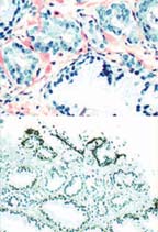
A 66 year old man with a PSA of 6.8 had a 12 quadrant biopsy of the prostate.
Histology
Core biopsy of the left lateral base shows a minute cluster of small acini with open lumina between hyperplastic prostatic glands.Immunophenotype
The small acini lack basal cell staining with P63 and high molecular weight cytokeratin 34BE12. Racemase (P504) show intense lumina stain.Diagnosis
Adenocarcinoma of prostate, Gleason's grade 3+3 (6), involving less than 1% of the tissue core. -
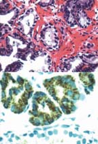
A 63 year old man had a 6 quadrant biopsy of the prostate. A diagnosis of benign prostate tissue was made by another laboratory. A second opinion from a CBLPath pathologist was requested.
Histology
A minute cluster of small acini with open lumina was seen in sections from the left base.
Immunophenotype
The acini lack basal cells as demonstrated by high molecular weight cytokeratin 34BE12 and with P63. P504 was positive.
Diagnosis
Adenocarcinoma of prostate, Gleason's grade 3+3 (6), involving less than 1% of the tissue core. -
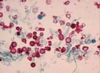
A 49 year old woman presenting with intermittent hematuria.
Specimen Type
First morning void.
Diagnostic Findings
Mild renal bleeding.
Diagnostic Significance
Hematuria (blood in the urine) is identified as either dysmorphic indicating upper urinary tract bleeding or isomorphic indicating lower urinary tract bleeding. Increased numbers of dysmorphic erythrocytes in the absence of additional pathologic findings is often associated with stress and exercise. Laboratory statistics reveal 79.38% of all urine cytology specimens submitted present with microhematuria with 28.35% presenting with mild renal (dysmorphic) bleeding. -
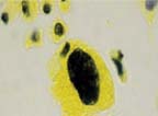
A 50 year old man presenting with asymptomatic hematuria.
Specimen Type
Voided urine.
Diagnostic Findings
Significant atypia present, moderate urothelial atypia.
Diagnostic Significance
Studies have shown (Laino, et al.) that classification of urothelial atypia is possible using a Feulgen staining technique and that 20% of all mild urothelial atypia progress to a more significant lesion within 2 years. Moderate urothelial atypia is a condition that may be associated with carcinoma in situ. Laboratory statistics reveal 4.26% of all urine cytology specimens submitted present with either mild or moderate atypia.