
Case Studies
-
A bone marrow biopsy is performed on a 76-year-old woman presenting with leukocytosis, thrombocytosis, and anemia.
Morphology
The bone marrow core, clot, and aspirate smears show megakaryocytic hyperplasia including many enlarged forms with hyperlobulated nuclei and abnormal nuclear:cytoplasmic ratios. Myeloid hyperplasia with maturation is also seen. There is no increase in blasts.Special Stains
A reticulin stain shows moderate fibrosis (MF-2).Flow Cytometry
Flow cytometry shows no evidence of a lymphoproliferative disorder and no increase in blasts.
FISH
FISH analysis is negative for BCR/ABL rearrangement.
Cytogenetics
An abnormal female karyotype including trisomy 8 is seen in 8 of 20 metaphases analyzed.
Molecular
CALR mutation is detected. JAK2 V617F and MPL mutations are not detected.Diagnosis
Primary myelofibrosis -
A core needle biopsy is performed on an enlarged cervical lymph node in a 69-year-old man.
Histology
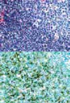
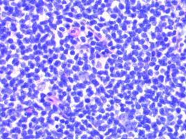
There is a diffuse and nodular proliferation of predominantly small lymphocytes with round to irregular nuclei, inconspicuous nucleoli, and scant cytoplasm.
HistologyImmunohistochemistry
The vast majority of the lymphocytes are abnormal B cells expressing CD20, CD5, BCL2, cyclin D1, and SOX11.
Flow Cytometry
An abnormal monotypic B-cell population expressing CD20, CD19, CD5, and lambda is detected.
Diagnosis
Mantle cell lymphoma