
Case Studies
-
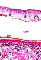
A 79 year old woman with an irregular, flat lesion, with shades of gray, brown and black on her temple.
Histology
Poorly circumscribed and asymmetrical melanocytic proliferation composed of cells arranged in solitary units and in nests along the dermoepidermal junction and focally above it. Individual cell shave clear to amphophilic cytoplasm with enlarged and hyperchromaticor vesicular nuclei, some with prominent nucleoli.
Diagnosis
In situ melanoma, lentigo maligna type, extending to the edges of the biopsy specimen. -
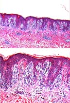
A 27 year old man with a well-demarcated pink nodule on his abdomen.
Histology
Well-defined, symmetrical melanocytic proliferation composed of regularly distributed nests along the dermoepidermal junction. Clefts are noted around the nests. The epidermis is hyperplastic and rare pink bodies are noted. Melanocytes are also seen in the dermis showing a normal maturation pattern of decreasing size in their progressive descent into the dermis.
Diagnosis
Compound melanocytic nevus, Spitz type. -
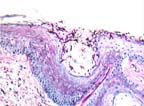
A 36 year old man with a scaly, red, flat lesion on his leg.
Histology
There is mild orthokeratosis and a minimal superficial perivascular lymphocytic dermatitis. Filamentous and budding yeasts, best seen with the PAS stain, are seen in the keratin layer.
DiagnosisSuperficial dermatophytosis consistent with Tinea species.
.
-
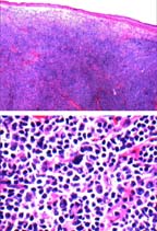
A 79 year old man with a scalp nodule of several months duration.
Histology
Dermal nodule extending into the subcutis composed of amixed population of small and large lymphocytes. The larger cells have vesicular nuclei and prominent nucleoli. Immunostain shows the larger cells staining with B-cell markers and the smaller cells withT-cell markers.
Diagnosis
Malignant lymphoma B-cell, T-cell rich cell type.