
Case Studies
-
A rectal polyp was removed from a 56 year old woman.
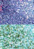
Histology
There is a submucosal mass giving a polypoid appearance to the lesion. The mass is composed of lymphoid nodules. A proliferation of small lymphocytes is seen outside the lymphoid follicles. There are well formed germinal centers and preserved mantle zones.
Immunophenotype
By immunohistochemical studies, large number of cells are B-cells showing expression with CD20 and CD79a. The B-cells are seen in the nodules and in the interfollicular area. The B-cells within the germinal centers are blc-6 and CD10 positive and show a fragmented follicular dendritic meshwork with CD21. These cells are bcl-2negative. IgD shows an irregular mantle zone. A few CD3 and CD5positive T-cells are present.
Diagnosis
Marginal zone lymphoma. -
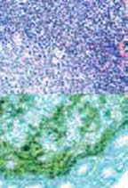
A rectal polyp was removed from a 21 year old woman.
Histology
Polypoid mass composed of lymphoid nodules with wellformed germinal centers and intact mantle zones.
Immunophenotype
The lymphoid cells show a mixture of T- and B-cells. There is an intact meshwork of follicular dendritic CD 21 positive cells. The follicular nodules are bcl-2 negative. The B-cell nodules are surrounded by CD3 positive T-cells. The plasma cells are polytypic.
Diagnosis
Benign follicular hyperplasia. -
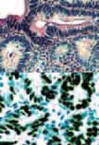
A 45 year old man had multiple colon biopsies for chronic ulcerative colitis surveillance. On endoscopy, the patient had a pan-colitis, most severe in the rectum. No mass was identified.
Histology
Mild to severe active colitis with chronicity were seen in several biopsies. In the rectum, the biopsy showed small glands arranged in aback-to-back pattern, lined by cells with elongated, hyperchromatic nuclei, some with prominent nucleoli, in a pseudostratified pattern. Numerous mitoses are seen.
Immunophenotype
Marked immunostaining for P53 and proliferating antigen Ki-67.
Diagnosis
High grade dysplasia/intramucosal adenocarcinoma in a background of ulcerative colitis.
Follow-up
Patient to undergo colectomy. -
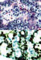
A 46 year old man had multiple esophageal biopsies for surveillance of Barrett's esophagus.
Histology
All biopsies show columnar mucosa lined by goblet cells. In one of the biopsies, the glands were smaller and the maturation process to the surface was disrupted.
Immunophenotype
The glands and surface epithelium show diffuse increased nuclear reactivity with the proliferating antigen Ki-67.
Diagnosis
Barrett's esophagus with low grade dysplasia.