
Case Studies
-
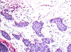
A 56 year old woman with a core biopsy of a palpable mass in the right breast.
Histology
Core biopsy showing nests of epithelioid cells arranged in irregular bands with poorly formed lumina.
Immunostain
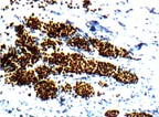
Immunostain for actin failed to detect myoepithelial cell saround the neoplastic glands. Immunostain for (a) Estrogen Receptor - 70% positive, (b) Progesterone Receptor - 70% positive and (c) Her 2Neu - 0/negative.
Diagnosis
Infiltrating moderately differentiated duct carcinoma of breast Estrogen and Progesterone Receptor Positive. -
A 41 year old woman with a lesion in the left breast at 3:00.
Cytopathology
There are 4 aspirate smears containing abundant epithelial clusters. They are variably sized and include many large aggregates, some with complex configuration. Cells within aggregates display moderate cytologic atypia and dyshesive tendency, with breakdown of large aggregates into small cellular clusters and occasional single intact cells.
Diagnosis
Significant findings present. This is a proliferative epithelial lesion with dyshesive tendency. Further evaluation, including histologic biopsy, is suggested for more precise classification of this lesion.
Follow-up
Based on the FNA results, the left breast 3:00 lesion was biopsied, and showed lobular carcinoma in situ.